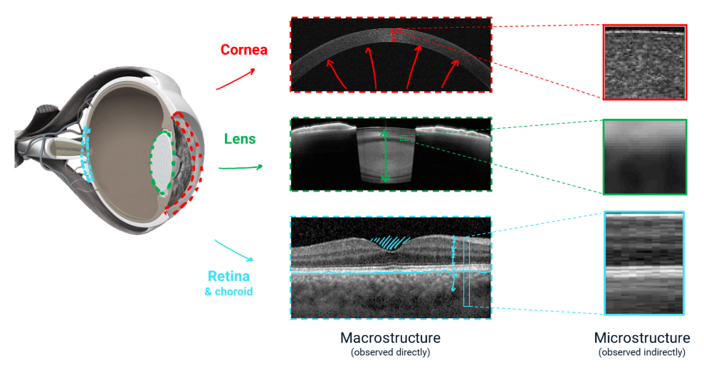
Clinical eye images could be explored in an entirely new way to provide early detection of eye diseases that currently affect thousands worldwide yet could be preventable. At VISIONSAFE, our objective is to extract macro- and microstructure biomarkers from real-world clinical images of the retina, lens, and cornea, thereby facilitating early disease detection and personalized management. While macrostructural biomarkers related to ocular shape are well-established, microstructural biomarkers related to tissue remain largely unexplored.
Our approach combines data science, advanced image processing, and wet-lab experimental work to better understand the interplay between macro- and microstructural biomarkers in prevalent sight-threatening eye diseases — such as, age-related macular degeneration (AMD), glaucoma, cataracts, and corneal ectasia — using efficient, cost-effective, and accessible tools.
At VISIONSAFE, we address several ground-breaking challenges: clinical eye data is already available but underused (can we use data science for vision preservation?); the acquisition and combination of macro- and microstructural biomarkers from clinical images have not yet been fully explored (is this combination a key for the early diagnosis of eye diseases? Are we missing out essential information from already available data?).
Research strategy
Our expertise integrates data science, clinical imaging, and experimental wet lab work.
Real-world
data
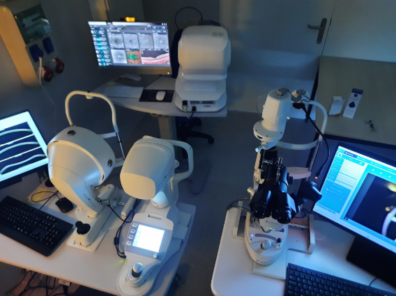
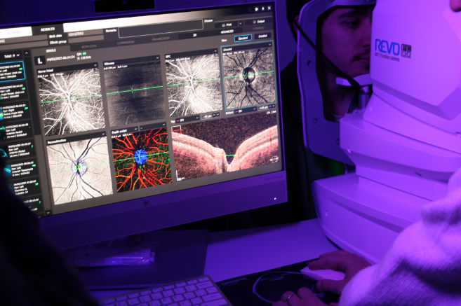
We collaborate with internationally recognized hospitals and universities for clinical data collection. Additionally, we are equipped with top-notch ophthalmic technology.
Macro and micro biomakers
We use image processing and artificial intelligence to reveal microstructural details in biological images beyond their resolution.
Multimodal imaging
We explore the synergy of combined imaging techniques to achieve better tissue characterization.
Disease screening
We evaluate the optimal combination of macro- and microstructure biomarkers for effective screening of each target eye disease.
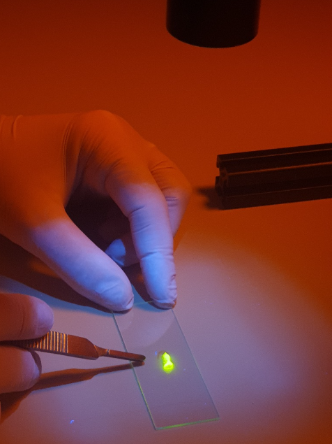
Experimental validation
We perform controlled tissue modifications in the cornea, lens, and retina ex vivo using high-resolution microscopy to link our data-analysis-driven biomarkers to real microstructural alterations.